


The five stages of diagnostic test process are very important to find the exact cause and determine appropriate treatment method. Through an accurate and precise diagnosis and treatment, we can increase the success rate of treatment by performing the most appropriate stage of treatment without sequelae and complications based on the patient’s condition.
1st Step

2nd Step
3rd Step

4th Step
5th Step
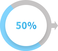
1st StepConsultation and X-rays
Diagnostic accuracy50%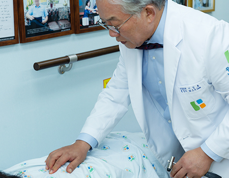
The initial diagnosis is made with the patient's medical history, symptoms, neurological examination, physical examination, and dynamic spine radiography, which shows the alignment of the spine.
Spinal x-ray is a very important examination in diagnosing the degree of degeneration of spinal bones, discs and joints, actual curvature, whether you have scoliosis or kyphosis, and instability of the spine during flexion and extension.
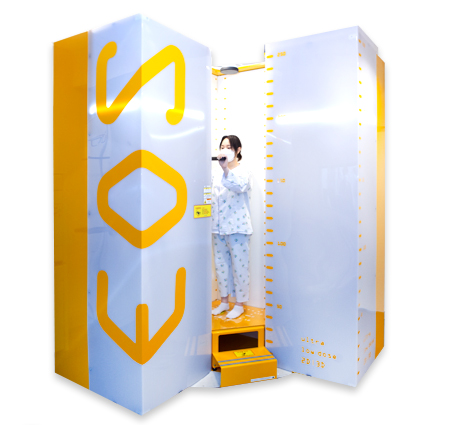
EOS X-ray Imaging System
EOS Imaging Scan is an innovative ultra-low dose 2D/3D X-ray imaging system.
An EOS Scan shows natural, weight-bearing posture and allows specialists to evaluate spinal alignment and interactions between the spine and the rest of the musculoskeletal system.
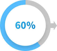
2nd StepMRI ScansTo check soft tissues, intervertebral discs and nerves
Diagnostic accuracy60%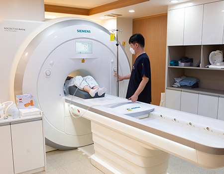
MAGNETOM Amira
Advantages of Wooridul’s MRI (Magnetic Resonance Imaging)
MRI uses a strong magnetic field and radio waves to generate images of parts of the body. MRI provides better soft tissue contrast and can differentiate between fat, water, muscle, and other soft tissue. Especially, spinal nerves, discs and soft tissue are clearly visible from the side.
3rd StepCT ScansTo check vertebrae, ligament calcification and spinal joints
Diagnostic accuracy70%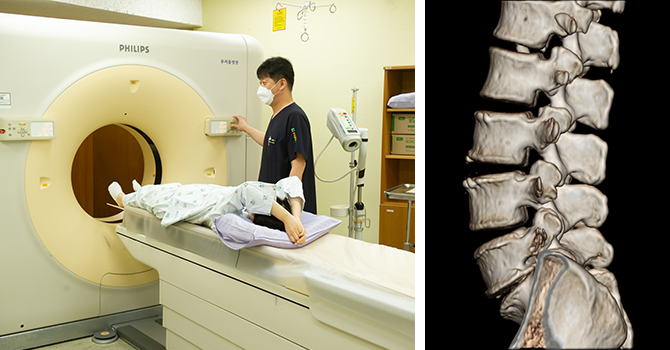
CT, Computed Tomography
CT uses specialized X-ray equipment to produce cross-sectional images or “slices” of the body.
It contains more detailed information than conventional x-rays.
CT provides clear images of spinal bones, soft tissue and calcified lesions.
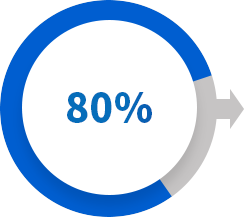
4th StepMR MyelographyTo check spinal cord and spinal canal
Diagnostic accuracy80%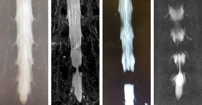
MR myelography uses a non-invasive magnetic resonance technique that allows specialists to evaluate the spinal canal and spinal cord without injecting a contrast agent into the subarachnoid space and shows the area where the nerve is compressed in detail.
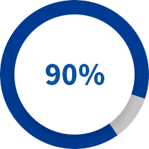
5th StepSpinal nerve function test
Diagnostic accuracy90%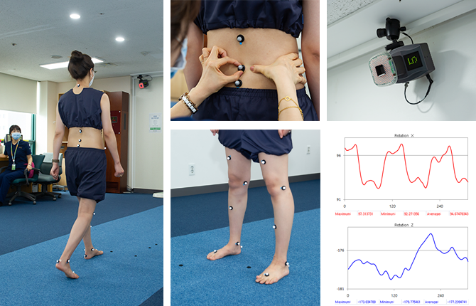
Spinal nerve function test
Spinal nerve function tests such as electromyography, motion analysis, and digital infrared thermal imaging can more accurately identify the lesion causing the patient’s symptoms. This can increase the accuracy of diagnosis and the success rate of treatment.
 Accreditation of Spine Specialty Hospital by the Korean Ministry of Health and Welfare
 Healthcare Accreditation by the Korean Ministry of Health and Welfare
 Healthcare Provider Certification for International Patients by the Korean Ministry of Health and Welfare
 Top 10 World’s Best Hospitals for Medical Tourists
 30 Most Technologically Advanced Hospitals in the World
 ‘World's Best Smart Hospital'
Copyright © 2026 Wooridul Spine Hospital
All Rights Reserved.

 Calling
 Appointment
 Our Doctors